endospore stain results|7.3: Endspore Procedures and Results : Clark Interpret results of an endospore stain. Identify when endospores are terminal, subterminal, and central in microscopic images, diagrams, and descriptions. Tell how the endospore stain works including the stains involved and how the stains penetrate cells . Video by PewDiePie: https://youtu.be/s0d-AFtXtkMSubscribe to my other Youtube channels for even more content! xQc Reacts: https://bit.ly/3FJk2IlxQc Gaming: h.

endospore stain results,Interpret results of an endospore stain. Identify when endospores are terminal, subterminal, and central in microscopic images, diagrams, and descriptions. Tell how the endospore stain works including the stains involved and how the stains penetrate cells .Learn how to stain bacterial endospores using malachite green and safranin or carbolfuchsin and nigrosin. See examples of positive and negative endospore stai.Endospore staining is a technique used in bacteriology to identify the presence of endospores in a bacterial sample. Within bacteria, endospores are protective structures used to survive extreme conditions, including high temperatures making them highly resistant to chemicals. Endospores contain little or no ATP which indicates how dormant they can be. Endospores contain a tough outer . Endospore staining is a differential technique that selectively stains the spores and makes them distinguishable from the vegetative .endospore stain resultsWith this staining procedure, the vegetative bacteria stain blue and the endospores are colorless. Note the "tennis racket" appearance of the endospore-containing Clostridium. .
7.3: Endspore Procedures and Results Endospore Staining is a technique used in bacteriology to identify the presence of endospores in a bacterial sample, which can be useful for classifying .
The Endospore stain, commonly referred to as the spore stain, is a type of staining technique. Its primary application is to locate and recognize the existence of . Learn how to detect and identify endospores and vegetative cells using different staining techniques, such as Schaeffer Fulton and Dorner methods. See the reagents, steps, results and applications of .Result: After staining the vegetative cells appear become colourless, the endospores stains as red which can be present as terminal or sub-terminal. The background is .endospore stain results 7.3: Endspore Procedures and Results Image 1: Endospore staining; a microscopic view of the cells being studied for. Picture Source: austincc.edu What is the significance of endospore staining? It helps in classifying and differentiating bacteria. .Endospore staining uses two stains to differentiate endospores from the rest of the cell. The Schaeffer-Fulton method (the most commonly used endospore-staining technique) . From the results of the Gram stain, the technician now knows that Cindy’s infection is caused by spherical, gram-positive bacteria that form grape-like clusters, which . INTRODUCTION TO ENDOSPORE STAINING. Some bacteria are capable of changing into dormant structures that are metabolically inactive and do not grow or reproduce. Since these structures are formed inside the cells, hence called endospores. These are remarkably resistant to heat, radiation, chemicals and other agents, that are .The LibreTexts libraries are Powered by NICE CXone Expert and are supported by the Department of Education Open Textbook Pilot Project, the UC Davis Office of the Provost, the UC Davis Library, the California State University Affordable Learning Solutions Program, and Merlot. We also acknowledge previous National Science Foundation support under .

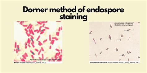
The all treatments staining have been effect on bacterial spores staining results. The warming time greatly affect the dye to penetrate the walls of bacterial spores, this can be . Schaeffer Fulton method that is widely used in painting endospores, endospore first stained with Malachite Green by a heating process, this solution is a powerful . Endospore staining history In 1922, Dorner published a set of staining instructions. He discovered a method of differential labeling called endospore stain that makes vegetative cells seem pinkish-red and endospores appear green under the light microscope.Dorner’s method included using heat as a stage, but because it required a .
Endospore staining is a crucial technique in bacteriology that identifies endospores within bacterial samples. . Related: Dorner method of endospore staining: procedure, principle, and results. Moeller stain technique. The Moeller staining technique, also known as the Moeller stain, is a method for staining endospores. This technique . Both the acid-fast and endospore stains require the use of steam to drive the dyes into bacterial cells (Figure below TBD). Due to time constraints, each student will perform one of the two staining methods. . The finer the mincing, the better your staining result will be. To prepare the smears for the endospore stain, add two drops of water . The Bacterial Endospore Stain on Schaeffer Fulton using Variation of Methylene Blue Solution. A Oktari 1, . 12), and varyous heating time (3, 4, and 5 minutes). The all treatments staining have been effect on bacterial spores staining results. The warming time greatly affect the dye to penetrate the walls of bacterial spores, this can be . The endospore coat is a multilayered shell that protects the bacterial genome during stress conditions and is composed of dozens of proteins. . mutations that disrupt the coat result in rapid .
The Malachite green staining (Schaeffer-Fulton method) is the most common method used to perform endospore staining.Malachite green stain can also used as a simple stain for bacterial cells.. The Schaeffer-Fulton method uses heat to push the primary dye (malachite green) into the endospore. Washing with water decolorizes the cell, but the endospore .The endospore stain is a differential stain used to visualize bacterial endospores. Endospores are formed by a few genera of bacteria, such as Bacillus . By forming spores, bacteria can survive in hostile conditions. .
The Schaeffer Fulton stain method also known as Wirtz Conklin Staining Method uses a combination of heat and chemical dyes to penetrate the tough walls of endospores and stain them green, while .
Endospore staining can define as one of the unique staining method, which is used to differentiate between the spore formers and non-spore formers by selectively staining the endospore within the vegetative cell. . which results in food spoilage and ultimately cause food diseases in humans and animals. Related Topics: Cyanobacteria .

Endospore stain of a Bacillus cereus culture using the Shaeffer-Fulton method and viewed at 1,000x total magnification under an oil immersion . so a negative result is not proof of a cell's inability to produce flagella. The demonstration slide was prepared with a silver compound to coat the flagella and a red dye, basic fuchsin, to . Acid-fast staining was developed by Robert Koch in 1882 and later modified by other scientists. Koch used the method to observe the “tubercle bacillus”—what we now call Mycobacterium tuberculosis, in sputum samples. While acid-fast and gram staining are both differential stains, the acid-fast stain is much more specific. The Interpretation of Endospore Staining Results. The Cells containing endospores appears as the red colored rod-shaped structure along with an intracellular spherical or elliptical green colored structure. It represents the red colored vegetative bacilli with green colored endospores (intracellular spores).
The endospore stain is a differential stain because it differentiates spore-formers from non spore-formers. Note: Formation of an endospore. The spore stains green and the vegetative cells stain red to orange. Procedure; Results; Lab 4 / Endospore Stain / Capsule Stain / Lab 4 Organisms. Please take a few .
endospore stain results|7.3: Endspore Procedures and Results
PH0 · Endospore staining
PH1 · Endospore Staining: Principle, Procedure, Results and Example
PH2 · Endospore Staining: Principle, Procedure, Results
PH3 · Endospore Staining: Principle, Procedure, Reagents, Results
PH4 · Endospore Staining
PH5 · Endospore Staining
PH6 · 7.3: Endspore Procedures and Results
PH7 · 2.2: Endospore Stain Procedure
PH8 · 1.12: Endospore Stain