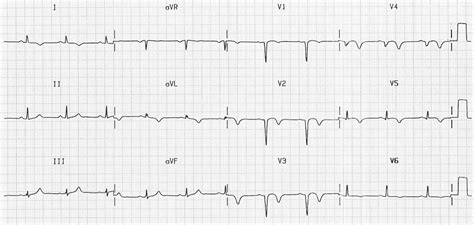
prwp ecg|ecg interpretation for nurses : Baguio Poor R wave progression is a common EKG pattern in which the expected increase of R wave amplitude in precordial leads does not occur 1. In a normal EKG, the . Find your ideal job at SEEK with 128 jobs found in Australia. View all our vacancies now with new jobs added daily! Gold coast city council Full Time Jobs in All Gold Coast QLD

prwp ecg,Poor R-wave progression (PRWP) is a common ECG finding that is often inconclusively interpreted as suggestive, but not diagnostic, of anterior myocardial .
Understand ECG basics. Medmastery; Wiesbauer F, Kühn P. ECG Mastery: .
ECG Library Basics – Waves, Intervals, Segments and Clinical Interpretation; .ECG Pearl. There are no universally accepted criteria for diagnosing RVH in . Poor R wave progression is a common EKG pattern in which the expected increase of R wave amplitude in precordial leads does not occur 1. In a normal EKG, the .Poor R wave progression (PRWP) is a common ECG finding that can indicate various cardiac conditions, such as ischemia, infarction, or ventricular hypertrophy. Learn the . CARDIOLOGY, ECG. Poor R wave progression (PRWP) refers to the absence of the normal increase in the size of the R wave in the precordial leads from .
Poor R-wave progression (PRWP) is a common clinical finding on the standard 12-lead electrocardiogram (ECG), but its prognostic significance is unclear. Poor R-wave progression as a .prwp ecg Poor R wave progression (PRWP) is a relatively common electrocardiogram (ECG) finding in adults, occurring in as many as 10% of all .Electrocardiographic “poor R-wave progression” is a troublesome clinical finding. Although commonly associated with prior anterior myocardial infarction, this finding is often seen in . Poor R-wave progression (PRWP) is a common electrocardiographic phenomenon in which the anticipated increase in R-wave amplitude in successive . The role of 12-lead electrocardiogram (ECG) in diagnosing myocardial infarction (MI) is well established , and poor R-wave progression (PRWP) is .Right ventricular parietal block, reduced QRS amplitude, epsilon wave, T wave inversion in V1-3 and ventricular tachycardia in the morphology of left bundle branch block are the .
In a series of 19,734 ECGs performed for a life insurance company and documented in the Journal of Insurance Medicine, PRWP was the sixth most common abnormal ECG pattern. 1 A survey of the . In a series of 19,734 ECGs performed for a life insurance company and documented in the Journal of Insurance Medicine, PRWP was the sixth most common abnormal ECG pattern. 1 A survey of the literature published in Cureus in 2020 concluded that the average incidence varies from 11.37% to 16.08% depending on the population .
Background: Poor or reverse R-wave progression (PRWP) is a common statement on electrocardiogram (ECG) interpretations, but its value in diagnosing anterior myocardial infarction (MI) is disputed. We assessed the accuracy of PRWP criteria in diagnosing anterior MI. Methods: We searched MEDLINE (1960-1998) and found 3 criteria for PRWP.Figure 1 Approach to patient with poor (PRWP) or reversed R-wave progression (RRWP). . In 13 patients with poor or reversed R-wave progression on the ECG and myocardial infarction of the left ventricular free wall at autopsy, 92 percent (12) had nontransmural myocardial infarction. In five patients with Q waves (QS, qR, or qr complexes) in .Background: Poor R-wave progression (PRWP) is a common clinical finding on the standard 12-lead electrocardiogram (ECG), but its prognostic significance is unclear. Objective: The purpose of this study was to examine the prognosis associated with PRWP in terms of sudden cardiac death (SCD), cardiac death, and all-cause mortality in general population .

ECG Library Function. LITFL ECG library is a free educational resource covering over 100 ECG topics relevant to Emergency Medicine and Critical Care. All our ECGs are free to reproduce for educational purposes, provided: The image is credited to litfl.com. The teaching activity is on a not-for-profit basis.prwp ecg ecg interpretation for nursesECG Library Function. LITFL ECG library is a free educational resource covering over 100 ECG topics relevant to Emergency Medicine and Critical Care. All our ECGs are free to reproduce for educational purposes, provided: The image is credited to litfl.com. The teaching activity is on a not-for-profit basis.ecg interpretation for nurses PRWP, atau Poor R Wave Progression per definisi adalah tidak adanya peningkatan ukuran gelombang R pada EKG dari V1 hingga V6, padahal semestinya ada. Atau mungkin sederhananya bisa dikatakan adanya kelainan dalam kelistrikan jantung. Memang untuk memahami ini sepenuhnya, dibutuhkan pemahaman terlebih dahulu .

執筆:Dr.ヒロ|杉山裕章. RV 3 ≦1mm:V 3 のR波高1mm以下 「R波の増高不良」(Poor R-wave progression)という所見は,言葉で表現してしまうと,「RV 3 ≦1mm」のようにシンプルでやや味気のないものに見えます。 ただ,字面通りに V 3 のr/R波「だけ」を見れば良いというものではありません 。
Abstract. Poor R-wave progression is a common ECG finding that is often inconclusively interpreted as suggestive, but not diagnostic, of anterior myocardial infarction (AMI). Recent studies have shown that poor R-wave progression has the following four distinct major causes: AMI, left ventricular hypertrophy, right ventricular hypertrophy, and . Background: The QRS pattern on the electrocardiogram (ECG) has an expected progressively increasing amplitude of the R waves in the anterior leads. Poor R wave progression (low R wave amplitude of < 3 mm in V3) is commonly reported, however its clinical significance is unknown. It may be related to underlying cardiac pathology, . Overview of Wolff-Parkinson-White (WPW) Syndrome. WPW Syndrome refers to the presence of a congenital accessory pathway (AP) and episodes of tachyarrhythmias. The term is often used . We examined the prevalence and prognostic impact of poor R-wave progression (PRWP) in a standard electrocardiogram (ECG) in a general population. Methods: The Health 2000 survey is prospective and nationally representative population cohort (random sample) health examination survey conducted in Finland in 2000–2001. . PRWP was the sixth most common abnormal ECG pattern in a consecutive series of 19,734 ECGs collected by the Metropolitan Life Insurance Company over a period of 5 ¼ years . Some studies suggest that 50% or more ECGs have lead misplacement errors which affects their analysis and interpretation [7,19,20]. Normal progression of the R wave in the precordial ECG leads implies transition between V 2 and V 3.The amplitude of the R wave in lead V 3 is usually greater than 2 mm. 1 A poor R wave progression (PRWP) has been defined as an absolute magnitude of the R wave less than or equal to 3 mm in lead V 3 and an R wave in lead V .
Poor R-wave progression (PRWP) is a common electrocardiographic diagnosis. However, the diagnostic usefulness of PRWP for coronary artery disease (CAD) and the plausible explanation for subjects with normal heart function are unclear. . (ECG) leads are determined by bony landmarks on the precordium. A measured R-wave .BACKGROUND Poor or reverse R-wave progression (PRWP) is a common statement on electrocardiogram (ECG) interpretations, but its value in diagnosing anterior myocardial infarction (MI) is disputed. We assessed the accuracy of PRWP criteria in diagnosing anterior MI. METHODS We searched MEDLINE (1960-1998) and found 3 criteria for .
Learn about PRWP ECG and how it can benefit your health. Nao Medical offers this service in NYC for your convenience. 1. Introduction. The role of 12-lead electrocardiogram (ECG) in diagnosing myocardial infarction (MI) is well established , and poor R-wave progression (PRWP) is interpreted as the probable anterior MI , .However, regeneration of R-wave or disappearance of Q-wave sometimes occurs after MI especially in the coronary .
prwp ecg|ecg interpretation for nurses
PH0 · steps to perform an ekg
PH1 · prwp medical abbreviation
PH2 · ekg interpretation practice
PH3 · ecg interpretation pdf
PH4 · ecg interpretation for nurses
PH5 · basic ekg strips and interpretation
PH6 · basic ecg interpretation powerpoint
PH7 · abnormal ecg interpretation
PH8 · Iba pa